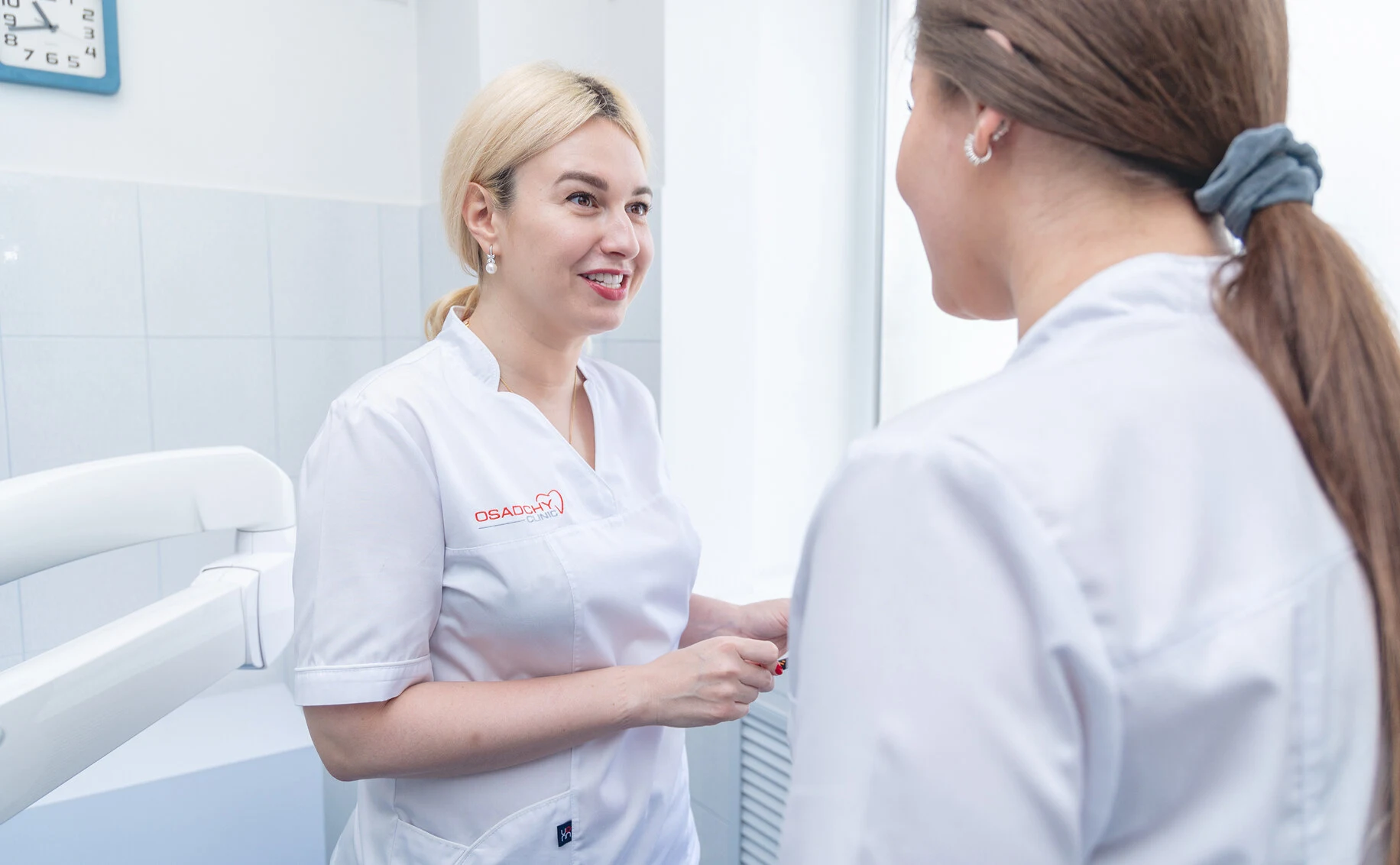
Didn’t find the necessary information? Make an appointment for a consultation with oral surgeon Ihor Oleksandrovych Osadchy.
Treatment of Teeth and Gums
Even minor pain or discomfort is already a reason to see a doctor for a consultation. As Hippocrates once said: “Pain is the watchdog of health.” That is why the sooner a patient responds to symptoms, the faster and more affordable the solution will be.


- a general dentist treats caries, periodontitis, pulpitis, periapical disease, and performs tooth restorations;
- a prosthodontist solves aesthetic and functional dental problems;
- an orthodontist corrects issues related to malocclusion and tooth alignment;
- an oral surgeon performs tooth extractions and dental implant placement, as well as other oral and maxillofacial surgery tasks.
The most common dental requests are treatment of gum diseases, caries, and pulpitis. Most clinics in Kyiv and Ukraine offer services for treating these conditions. However, before choosing one specialist over another, patients should familiarize themselves with certain nuances and details. Quite often, such knowledge helps avoid confusion and get quality dental treatment at an optimal price.
All dental diseases can be roughly divided into three groups:
- diseases of the crown portion;
- diseases of the root portion;
- diseases of the soft tissues surrounding the tooth (mucosa).
Each disease has its own treatment protocol that the doctor must follow. In this article, we will examine in detail the diseases that mainly fall within the competence of general dentists.
- Diagnosis of oral cavity diseases;
- Treatment plan development;
- Caries treatment;
- Pulpitis treatment;
- Periodontitis treatment;
- Periodontal disease treatment;
- Treatment of stomatitis and gingivitis;
- Endodontic treatment of chronic periapical tissue diseases;
- Tooth restoration;
- Professional oral hygiene;
- Teeth whitening.
- Pain in the oral cavity;
- Bleeding of soft tissues (gums);
- Halitosis (bad breath);
- Changes in tooth color;
- Increased tooth sensitivity (reaction to sweet, sour, cold, hot);
- Mechanical tooth damage (cracks, chips, injuries);
- Recession (exposed tooth root);
- Prevention (routine visits every 3–6 months).
Caries is a complex process that develops slowly in the hard tissues of teeth. As a rule, the disease begins asymptomatically and is difficult to detect except during a dental appointment.
The development of caries leads to damage of the hard tissues of the tooth with the formation of a cavity in the dentin (soft tooth tissue), and without treatment, it leads to inflammation of the pulp and periodontium — that is when painful sensations begin. Complications may also arise. It should be remembered that in an unfavorable course of caries, there is a high probability of tooth loss.
In summary: caries belongs to diseases of the crown portion of the tooth and is associated with the destruction of enamel and dentin tissues under the influence of acid processes.
Possible symptoms of caries:
- pain or tooth sensitivity;
- light, dark, or brown spots on the surface of teeth;
- discomfort while chewing.
Types of caries:
- superficial (initial enamel defects);
- medium (damage to deep enamel layers and partially dentin);
- deep (enamel damage and deep dentin damage).
Depending on the type, the following types of caries treatment are possible:
- filling (removal of damaged tissues and replacement with filling material);
- endodontic treatment (applied when the pulp is affected and involves canal filling).

Caries treatment becomes more complicated if the destruction is located close to the pulp. Placing a filling can lead to significant sensitivity and discomfort. This is because the pulp produces odontoblasts and forms dentin. Over time, the pulp becomes denser and smaller — it builds tissues around itself, essentially “walling itself in.” That is why, when treating deep caries, it is necessary to ensure that the pulp continues to produce dentin.
To achieve this, the doctor places a calcium-containing liner between the filling material and the pulp, which helps the pulp produce secondary dentin, thus keeping the tooth alive and nourished.
The use of a calcium-containing liner does not significantly increase the cost of caries treatment but greatly improves its quality. The patient does not experience discomfort after filling.
Pulpitis belongs to diseases of the root portion of the tooth, and its treatment involves:
- devitalization of the tooth (nerve removal);
- treatment of canals with a special antibacterial solution (and/or laser treatment);
- canal filling (using an apex locator to ensure that the filling material does not extend beyond the root tip).
Many patients fear depulpation, believing that by “killing” the nerve, the entire tooth is “killed” as well. Outdated methods, which are practically no longer used in Kyiv clinics, involved the use of aggressive materials. Indeed, under the influence of these materials, the tooth became more fragile. However, modern materials have become gentler, and a depulpated tooth covered with a crown can last quite a long time.
Typically, the cost of pulpitis treatment is influenced by the complexity of the case, the number of root canals in the given tooth, the amount of material used, and additional equipment.
At “Osadchiy Dental Clinic,” we use state-of-the-art endodontic equipment from leading manufacturers such as MicroMega and Parkell for root canal treatment. This allows us to perform a complete root canal treatment in just one visit without pain and prepare them for the next stage of filling with endodontic pastes.

Extensive experience
Стаж работы челюстно-лицевой хирурга 38 лет, из них 8 лет — работа дежурным врачом отделения острой травмы.

Number of surgeries
Over 2,000 successful surgeries in the field of maxillofacial surgery.

Quality of work
Thanks to a thorough approach in diagnostics and treatment planning for each clinical case, we achieve a minimal number of complications.
A microscope in dentistry is auxiliary equipment for the doctor that greatly facilitates their work. At the same time, the use or non-use of a microscope during treatment is not a factor that affects the outcome.
The use of an apex locator, which prevents going beyond the root tip, X-rays, and caries markers are important factors that significantly affect treatment outcomes, but the same cannot be said about the use of a microscope. It is necessary to reduce the strain on the doctor’s eyesight, but not to improve patient treatment results.
Most often, when it comes to dental treatment under a microscope, it means that the doctor will use a microscope for, say, faster detection of the canal entrance during pulpitis treatment. Finding the entrance to obliterated (narrowed) canals can be quite difficult, and significant magnification needs to be applied. At this stage, one can speak of dental treatment under a microscope with a slight stretch.
The fact that many dental clinics in Kyiv emphasize the use of a microscope speaks more about marketing than medicine. Catchy headlines like “Dental treatment under a microscope” allow raising service prices, but the quality of services will depend not on auxiliary tools, but on the experience and skills of the doctor. At “Osadchiy Dental Clinic,” we also use a microscope, but only when necessary, as the use of additional equipment increases the cost of dental treatment.
The commonly used word “gums” in dentistry is technically incorrect — the oral cavity contains mucous membrane. However, phrases like “gum diseases” and “gum inflammation” have become so firmly established that we are compelled to use this imprecise term.
Gum disease on its own is very rare; it is associated with an increase in conditionally pathogenic microflora. Sometimes it is provoked by physical, chemical, or microbial factors.
Types of mucosal diseases
| Name | Description |
| Stomatitis | Inflammation of the mucous membrane of bacterial, viral, or fungal origin. Manifests as white or reddish ulcers that cause itching and erosion of the cheeks, tongue, and gums |
| Glossitis | Inflammation of the tongue resulting from trauma, burn, or pathogen exposure. Causes a burning sensation; the tongue acquires a bright red color |
| Cheilitis | A lip disease caused by chronic skin conditions, sun, wind, or allergen exposure. Accompanied by peeling and the appearance of cracks in the corners of the mouth |
| Oral leukoplakia | Keratinization of the mucous membrane under the influence of aggressive factors (smoking, inadequate prosthetics, etc.). Appears as white, gray, or red plaques on the mucosa |
| Gingivitis | Inflammation of the gums due to poor hygiene. Affects only the surface of the gums. Accompanied by bleeding, gum swelling, pain or discomfort when pressing |
| Periodontitis | Inflammation of the periodontal tissues. Causes gum bleeding and exposure of tooth roots |
| Periodontal disease | A chronic disease of the tissues surrounding the tooth, non-inflammatory in nature |
Treatment of gum diseases depends on the type of disease. Local medication treatment, systemic treatment, surgical treatment, and combined treatment are used.
It should be noted that in 95% of cases, gum disease occurs as a result of a tooth disease. It is the reaction of surrounding tissues to an infection located in the tooth. In these cases, treatment of gum inflammation involves eliminating the source of infection in the tooth. Once the tooth is treated, the mucosa will also recover, most often without additional treatment of gum inflammation.
Supportive therapy is also possible. It involves biorevitalization of the oral mucosa, when medications that promote improved blood supply to tissues are administered. This can help suppress the inflammatory process.
There are many reasons for the development of tooth decay. The main ones include: insufficient attention to oral hygiene; specific saliva composition that results in inadequate self-cleaning of teeth; smoking and drug use; taking antibiotics during childhood when permanent teeth are forming; excessive consumption of high-sugar products.
When the tooth nerve is inflamed, the pain is so severe that not everyone can endure it. But even if you have started taking painkillers, sooner or later the pain will return, and the consequences will be much worse — the nerve inflammation will progress to a new level, where pulpitis transforms into periodontitis (inflammation of the bone tissue around the tooth root) or chronic tooth nerve inflammation.
Tooth decay itself is not critically dangerous; its complications are dangerous. The most common complication is pulpitis — inflammation of the tooth nerve.
A tooth chip is also possible with massive caries damage. The walls of the tooth become thinner and can break off even under light chewing pressure.
Dental treatment under general anesthesia can be performed, but we do not recommend it to our patients. It is important to remember that dental treatment involves several appointments, and using general anesthesia each time is quite dangerous. The use of anesthesia is primarily beneficial for clinics, as the anesthesia itself and its maintenance cost money. All of this significantly increases the cost of dental treatment. At our clinic, we use general anesthesia only when there are direct medical indications.
Signs of tooth nerve inflammation include unprovoked pain that significantly worsens at night. Tooth pain persists long after the irritant is removed. Often during pulpitis, the patient cannot tell exactly which tooth hurts; the pain can radiate to the teeth of the opposite jaw, to the ear area, and even to the eyes.
In cases of periodontitis and periodontal disease, when there is infection in abnormally deep periodontal pockets, vector therapy can be used. It involves the doctor using ultrasound to enter the pockets, clean them with a special disinfecting solution, and polish the tooth neck. Vector therapy helps delay the progression of the disease but does not cure it completely. At our clinic, we use vector therapy when medically indicated.
When visiting a dentist, it is advisable to have an allergy test for anesthesia, after which the doctor will administer a painkiller into the gum. After some time, the doctor will open the tooth cavity where the nerve is located, treat the canals (remove the nerve, clean the canal walls, rinse the canal and sterilize it). Then canal filling is performed and a follow-up X-ray is taken.
On our website you will find the basic price list for all our services, including caries treatment. The price may vary depending on the complexity of the case and the need to use additional equipment.


 Reviews

Ой ,як добре ! Зуби як в молодого! Щиро дякую та бажаю більше вдячних клієнтів. Імплантами задоволений на 100%, а лікуванням зубів ще більше.
Дай бог Вам процвітання а Ігорю Олександровичу довгії літа і мою повагу.
Володимир.
Read more

Добрий день. Хочу поділитися своїми враженнями про стоматологію. Пролікувалися всією родиною, залишилися дуже задоволені! Хороше сучасне обладнання та технології. Лікарі дуже ввічливі, знають свою роботу на всі 100 процентів. Дуже уважний, професійний лікар Шляховий Олексій Анатолійович, завдяки йому не так страшно йти до стоматолога))). Дякуємо за якісне та безболісне лікування! Рекомендую всім!
Read more

Дуже комфортна та професійна клініка! Вперше звернувсч до пані Костенко Наталії Миколаївні 12 років тому, і протягом усього часу жодного разу не пошкодував. Задоволений професіоналізмом лікаря, якістю послуги та вмінням знайти індивідуальний підхід до кожного клієнта. Послугами клініки користується вся наша родина. Тому із задоволенням рекомендую! А лікарям клініки бажаю успіхів, розвитку та усміхнених клієнтів!
Read more

Дякую за Вашу роботу! Гарна і охайна клініка, ввічливий персонал. Звернулась до лікаря Катерини Олександрівної для встановлення імпланта. Перед цим мені запропонували зробити чистку і вилікувати карієс. Роботою дуже задоволена, буду звертатись у майбутньому і рекомендувати друзям і знайомим!
Read more

Щойно повернулася з прийому в стоматології – і я під враженням! 🙌
Все пройшло супер комфортно: привітні адміністратори, сучасне обладнання та головне — уважний лікар, який пояснив і зробив акуратно і без болю.
Пішла з усмішкою, і тепер точно знаю, куди повернуся наступного разу. Дякую вам за турботу та атмосферу! 💙🦷
Read more

Відвідуємо стоматологію всією родиною.
Ключовий фактор це компетентність спеціалістів.Шляховий Анатолій Олексійович допоміг вирішити проблему, яка дошкуляла не один рік. Щиро дякуємо!
Катерині Олександрівній, своїм підходом до роботи з пацієнтом позбавила страху стоматологів))
Професіоналізм та сервіс заслуговують найвищої оцінки❤️
Read more

Я залишилась дуже задоволена! Атмосфера приємна, все чисто й затишно. Процедура пройшла швидко та максимально комфортно. Особливо дякую Осадчому Ігорю, все робить акуратно й професійно, а ще підтримує теплу розмову — час пролітає непомітно 😊
Read more

Завжди лікусь лише у Самокіної Катерини Олександрівни, дуже рекомендую, чудовий лікар + зручна локація клініки, все подобається.
Read more

Дуже хороша клініка, зокрема лікар Наталія Миколаївна 🤍 проходила у неї лікування брекетами, і досі настільки задоволена результатом, що і тепер чоловік теж проходить тут лікування. Завжди приємне ставлення 🫶🏼
Read more

Дякую Катерині Олександрівні за чудову роботу! Справжній професіонал з “золотими руками”. Навіть найскладніші процедури проходили абсолютно безболісно – у неї дуже легка рука.
Read more

Про лікаря Ігоря Олександровича дізналися завдяки YouTube, і це було найкраще рішення звернутися саме до нього. Він проводить установку імплантів без синус-ліфтингу – складну роботу, за яку більшість лікарів не беруться.
Щиро вдячні за його професіоналізм, уважність і людяність. Весь процес проходив комфортно та безболісно. Атмосфера в клініці дуже приємна, персонал доброзичливий, відчувається турбота й щире бажання допомогти.
Завдяки досвіду та сучасному підходу лікаря отримали чудовий результат без зайвих процедур.
Відтепер із упевненістю довіряємо здоров’я зубів саме Ігорю Олександровичу!
Окрема подяка ❤️ керівнику клініки Діані! За оперативне реагування на нашу проблему та бажання допомогти!
Ви команда професіоналів!
Read more

Рекомендую 100%
Скористалась лише однією послугою – панорамний знімок щелепи, але вже можу оцінити ставлення до клієнта та сервіс в загальному. Люди працюють на високому рівні це точно.
Read more

Клініка дуже гарна. Дизайн розслабляє та знімає напругу перед та після відвідування лікаря. Персонал клініки попереджувальний та уважний, з перших хвилин відчуваєш, що ти їм дорогий, вони раді тебе обслужити. Про лікарів можу сказати, що перший раз у свої 67 років, на прийомі навіть трохи подрімала. Це ж нонсен … але це так. Рекомендую
Read more

Вже не вперше звертаюся до цієї клініки – все завжди дуже швидко та якісно. Окрема подяка Осадчому Ігорю за професіоналізм і уважне ставлення: видалення зубів пройшло швидко, безболісно та комфортно!
Read more

Дуже рекомендую!
Робив повний цикл: лікування зубів, видалення мудрих, установка брекетсистеми, потім відбіл. Чудово!
Дружина видаляла відразу 4 зуби мудрості під наркозом – все було супер та швидке відновлення!
Доньці зубки теж лікували ⭐️👍
Read more

Нам дуже сподобалося. ПРИЙНЯЛИ дитину без запису попереднього адже болів зуб. Дякую🫶🏻Сервіс на вищому рівні, працюють професіонали своєї справи.
Read more